 |
Calcium |
 |
Tissue Distribution of Ca
Physiological Role of Ca
Absorption and Metabolism of Ca
Ca Homeostasis
Interaction of Ca with Other Dietary Components
Ca Requirements
Deficiency of Ca
Ca Excess
I. Tissue Distribution of Ca
- 99% of the body's Ca is in bones and teeth
- The calcium phosphate of bone is deposited within a soft fibrous organic matrix of collagen fibers, and to a much lesser extent, in a mucopolysaccharide gel
- Bone mineral consist of two calcium phosphate phases:
- Amorphous or nonchrystalline phase, a hydrated tricalcium phosphate
- A crystalline form, resembling hydroxyapatite
- Young bone contains more of the amorphous phase which is the precursor of the apatite phase
- Mature bone contains more of the apatite phase
- 1% of the body's Ca is outside the bone where it functions in a number of essential processes
- In serum, 60% of the Ca is ionized and physiologically active
- A significant decrease in serum ionized Ca results in tetany
- An increase can cause cardiac or respiratory failure
- The remaining serum Ca is nonionized and physiologically inert
- 35% of serum Ca is bound to protein
- 5% is complexed with citrate, bicarbonate, and phosphate
II. Physiological Role of Ca
- Essential for bone and tooth formation and maintenance
- Structural or supporting component of the body
- Readily available source of Ca for maintenance of homeostasis
- Required for normal blood clotting
- Ca must be present for prothrombin to form thrombin (EDTA or citrate function as anticoagulants by chelating Ca++ so it is unavailable to react the prothrombin)
- Thrombin reacts with fibrinogen to form the blood clot, fibrin
- Ca2+ plays a major role in the function of the nervous system
- Calmodulin is a major Ca2+-binding protein, which regulates many of the neural functions of Ca++ (see Fed. Proc. 41:2265.1982)
- Secretion and biosynthesis of monoamine neurotransmitters is stimulated by Ca++ influx in the nervous system which is regulated by calmudulin-dependent protein kinase II
- Ca++ is necessary for contraction of skeletal, cardiac, and smooth muscle (See Fed. Proc. 41:2223-2250 and 2863-2904.1982)
- Ca++ is important along with K+ and Na+ ions in the regulation of heartbeat
- Ca++ can act either as an activator or stabilizer of enzymes (Fed. Proc. 41:2253-2257.1982)
- Ca2+ is necessary for the secretion of a number of hormones and hormone-releasing factors (Fed. Proc. 2278-2282.1982)
- Ca plays a critical role in insulin release from the pancreatic b cell (Fed. Proc. 41:2278-2282.1982)
- The major physiological stimulus for insulin secretion is the concentration of glucose in extracellular fluid (Insulin secretion is also stimulated by elevated extracellular K+)
- Effects of glucose are closely linked with stimulation of electrical activity associated with alterations in influxes of various ions, particularly Ca2+
- Glucose stimulates Ca2+ influx and inhibits Ca2+ efflux from the b cell
- Resulting increase in cellular Ca2+ stimulates insulin release mediated by calmodulin-dependent protein kinase
- cAMP also triggers insulin secretion mediated by cAMP-dependent protein kinase
- Ca++ is required for activation of K+ channels
- Ca is involved in integrity of intracellular cement substances and membranes
- Calmodulin (Fed. Proc. 41:2251-2299.1982) is the major intracellular Ca2+ receptor (Also see J. Dairy Sci. 70:1551.1987)
- Calmodulin is a ubiquitous Ca2+-binding protein
- The major intracellular Ca2+ receptor
- Molecular weight 16,700 daltons
- Contains 4 Ca2+ binding sites
- It has no intrinsic enzymatic activity but it regulates a wide spectrum of enzymes that control many basic cellular processes
- Metabolism of Ca2+ in the cell
- Calmodulin activates membrane Ca2+ transport by activating (Ca2+ -Mg2) – ATPase
- Contractile activity and stimulus-secretion coupling – In the electric eel, 2% of the total protein is calmodulin
- Metabolism of cyclic nucleotides – calmodulin is the activator of cyclic nucleotide phosphodiesterase (see J. Dairy Sci.70.1551.1987)
- Mode of action of calmodulin (Fed. Proc. 41:2253.1982)
- Ca2+ that flows in or is released from the cell membrane or internal organelles bind to calmodulin
Ca2+ + Calmodulin --------------- Ca2+ • Calmodulin
(inactive) (active)
- Binding of Ca2+ to calmodulin induces a conformational change able to interact with phosphodieterase to form active complex
Apoenzyme + Ca2+ • Calmodulin -------- Ca2+ • Calmodulin • Enzyme
(less active) (active) (activated)
III. Absorption and Metabolism of Ca (J. Dairy Sci. 69:604:1986)
- Ca is absorbed primarily from the proximal small intestine by an active transport mechanism
- Facilitated transport of Ca across the duodenal brush border is initiated by 1,25-dihydroxy-vitamin D
- 1,25(OH)2D enters the enterocytes by diffusion and binds to its receptor in the cytosol (Enterocyte = intestinal cell)
- The 1,25(OH)2D-receptor complex translocates to the chromatin fraction of the enterocyte nucleus
- Synthesis of messenger RNA and specific proteins that control Ca transport is increased
- Ca transfer within the cytoplasmic compartment of the enertocyte:
- May involve vitamin D-dependent Ca-binding protein or encasement of Ca into vesicles
- Several organelles, mitochondria, endoplasmic reticulum, and the golgi sequester Ca and prevent its buildup within the cytoplasm
- Ca is excreted primarily via the feces
- The extracellular Ca pool is regulated within narrow limits by three hormones:
- Parathyroid hormone secreted from the parathyroid
- 1,25-dihydroxy vitamin D produced in the kidney
- Calcitonin secreted from the ultimobranchial bodies of the thyroid gland
- Bone metabolism of Ca and P
- Bone formation
- Endochondral bone formation in young animals. In young animals, bone formation occurs as a result of calcification of a specialized organic matrix
- The epiphyseal plate cartilage elongates by proliferation of the resting chondrocytes
- These cells prepare their organic matrix for mineralization
- Mineral is deposited on the matrix to give rise to endochondral calcification
- Intramembranous bone formation
- Does not involve chondroblast-mediated calcification
- Instead, osteoblasts form a membrane which elaborates organic matrix
- Mineral is deposited on the membrane
- Shaping of bone involves a combination of endochondrial and intramembranous formation
- Vitamin D plays an intimate role in both mineralization processes
IV. Ca Homeostasis
 |
- Ca in plasma is in exchange with a pool about 35 times larger than the amount circulating in blood
- Supply of Ca to the pool
- Intestinal absorption
- Bone resorption
- Exit of Ca from the pool
- Feces
- Urine
- Deposition in bone
- Developing fetus in pregnant female
- Mammary gland in lactating female
- Maintenance of the extracellular Ca pool depends on a balance between entry and exit of Ca from the pool
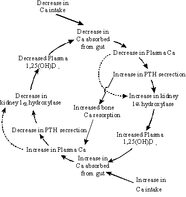
- Hormonal regulation of the extracellular Ca pool
- A slight decrease in serum Ca causes an increased secretion PTH
- PTH stimulates biosynthesis of 1,25-dihydroxy vitamin D which increases absorption from the intestine and Ca resorption from bone
- A slight increase in serum Ca results in a decrease in PTH secretion and an increase in calcitonin release
- These changes decrease production of 1,25(OH)2D and reduce intestinal absorption and bone resorption of Ca
 |
Mechanism of Adaptation to Alterations in Dietary Ca (Littledike and
Goff 1987. J. Anim. Sci. 65:1727-1743.) (Horst 1986. J. Dairy Sci.
69:604-616) (Dashed line represents a response in rats but not
in ruminants) |
- Bone mobilization in support of plasma Ca and P concentrations
- Both 1,25(OH)2D and PTH are needed for bone resorption
- Calcitonin lowers circulating Ca and P levels
- It exerts its Ca-lowering effect by inhibiting bone resorption
- This action is apparently due to inhibition of Ca++ permeability of osteoclasts (multinuclear cells that erode and resorb previously formed bone) and osteoblasts (bone-forming cells that secrete collagen, forming a matrix around themselves which then calcifies)
- Calcitonin also decreases osteoclast activity and number
- Calcitonin appears to be relatively inactive in adult humans and animals
- The hormone is more active in young individuals and may play a role in skeletal development
- It may protect the bones of the mother from excess Ca++ loss during pregnancy
- Bone formation in the infant and lactation are major drains on Ca++ stores
- Plasma concentrations of 1,25(OH)2D and calcitonin are both elevated in pregnancy
V. Interaction of Ca with Other Dietary Components
- Excess dietary phosphorus accelerates bone resorption
- 1. Ca to P ratio lower than 2:1 enhances bone resorption at both high (1.2%) and low (0.6%) Ca intakes
- 2. Thus bone loss in adult animals is achieved by either excess dietary P or insufficient dietary Ca
- Urinary loss of Ca in humans increases with rising dietary protein intake
- Phytates and oxalates may decrease Ca absorption by combining with Ca to form insoluble salts within the intestinal lumen
- Certain amino acids and lactose may enhance Ca absorption
- Hypomagnesemic animals often also become hypocalcemic
- There is no evidence hypomagnesemia reduces absorption of Ca
- Correction of the hypocalcemia depends on restoring Mg
- Infusion with Ca increases serum Ca only as long as the infusion lasts
- When Mg concentrations are restored, the hypcalcemia, after a lag period, returns to normal
- There appears to be a failure of the PTH mechanism at two levels in maintaining Ca homeostasis
- Sometimes, but not always, a failure to elevate PTH in response to low serum Ca
- A failure to mobilize bone Ca when PTH is elevated
- Impaired response of PTH during Mg deficiency may be due to essentiality of Mg to cyclic AMP
- Cyclic AMP mediates the action of PTH on bone and renal cortex
VI. Ca Requirements
- The amount of Ca required under various dietary conditions is not clear
- The confusion is probably due to the many factors – protein, P, F, hormonal factors – which appear to influence metabolism of Ca or mineralized tissues
- Recommended allowances of Ca (% of DM):
| Ruminants |
Ca |
P |
| Baby Calves |
0.6-.7 |
0.4-.5 |
| Growing heifers and bulls |
0.3-.5 |
0.2-.3 |
| Dry pregnant cows |
0.37 |
0.26 |
| Lactating cows |
0.4-0.65 |
0.31-0.38 |
| Mature bulls |
0.24 |
0.18 |
| Nonruminants |
Ca |
P |
| Starting chicks (0-8 weeks) |
0.9 |
0.7 |
| Growing chickens (8-18 weeks) |
0.6 |
0.4 |
| Laying hens |
3.25 |
0.5 |
Growing and finishing swine
1 to 5 kg
5 to 10 kg
10 to 20 kg
20 to 35 kg
35 to 60 kg
60 to 100 kg
|
0.9
0.8
0.65
0.6
0.55
0.5
|
0.7
0.6
0.55
0.5
0.45
0.4
|
| Breeding swine |
0.75 |
0.6 |
VII. Deficiency of Ca
- The primary symptom of Ca deficiency of young animals is rickets
- Rickets can be caused by a deficiency of Ca, P, or Vitamin D (for clarity, the terms low-Ca, or low P rickets should be used)
- Ca and P are not deposited in the organic cartilaginous matrix in sufficient quantities to develop a strong, dense bone
- Swollen tender joints
- Enlargement of bone ends, and bending of ribs
- Beading of ribs
- Arched back and stiffness of legs
- Structural abnormalities
- Buckled knees
- Bowed or crooked legs
- The weight of the body and tension of growing muscles pull the weakened, soft bones out of shape
- Osteomalacia is a decrease in mineral content of adult bone
- Caused by lack of adequate Ca and/or P in the diet
- Even when bone is mature, there is a continuous mobilization of Ca and P which must be replaced
- Pregnancy and lactation are periods of high Ca and P demand so mineral content of bone may be depleted
- Continual depletion of Ca and P from adult bone will lead to weak and brittle bones which may break under external pressure (when bone resorption exceeds bone deposition)
- High P intake can also cause osteomalacia
- Osteoporosis in man denotes the result of a defective synthesis of the protein matrix of bone
- Commonly occurs in old age, with a resulting decalcification
- Bone loss may be sufficient to cause disabling symptoms
- Several factors, including Ca intake, are probably involved
- Inactivity can lead to bone demineralization (astronauts during weightlessness in outer space)
- Milk fever (parturient paresis) (J. Dairy Sci. 69:604.1986)
- A sudden drop in blood Ca results from formation of colostrum
- Approximately 2.5g Ca are required for each kg of colostrum produced
- This is about the total amount of Ca in the blood at any given time
- A cow yielding 25kg of milk must replace her total blood Ca every hour
- A fall in blood Ca normally stimulates increases of plasma PTH and 1,25(OH)2D
- Increased bone resorption
- Increased intestinal Ca absorption
- Milk fever prone cows fail to respond to these stimuli
- Unable to mobilize Ca from bone rapidly enough
- Plasma Ca falls to critically low concentrations
- Coma and death may result
- Prevention of milk fever
- Feed low Ca diet before parturition to initiate Ca mobilizing mechanisms
- Increase anion, but not cation, content of prepartum diet
VIII. Ca Excess
- The homeostatic mechanism tends to protect against the absorption of excessive quantities of Ca
- Because of the interrelationships with other nutrients, especially P, excessive Ca for extended periods can harm animal performance
- Optimum animal performance is linked closely with Ca and P levels in the diet
- Most animals require a fairly narrow Ca to P ratio (usually no wider than 2 to 1)
- Ruminants can tolerate wider ratios than monogastric animals providing dietary P is adequate
- Excessive dietary Ca causes hypercalcitoninism in bulls (Krook et al.1971. Cornell Vet.61:625)
- Occurs in bulls fed excess Ca
- Excess Ca intake causes hypersecretion of calcitonin
- Normal bone resorption is inhibited and bone mass increases
- Bulls rarely have a need to resorb bone (as opposed to lactating cows)
- Osteopetrosis occurs where bone is deposited more rapidly than it is resorbed over a long time
- Pathological tissue decalcification – Ca is abnormally deposited in soft tissues in Mg deficiency

 For individual consultation or questions about our products, call
1-800-628-0997

